

![]()
![]()
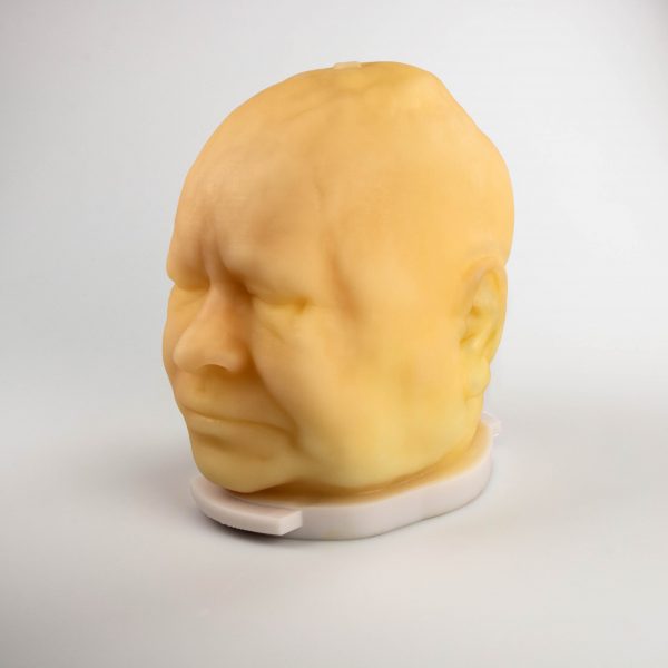
Biomimetic medical models include representation of major anatomical structures like bone, muscle, fat, vasculatrue, connective tissues, and skin. The 3D Systems ProJet/5600 printer with blended material inks and melt away wax support is used to print each optimized structure. These advanced anatomical models provide realistic feedback ideal for use as a cost-effective training tool for medical professionals and as a surrogate for advanced benchtop testing.
The focus of this project is the development of a technical data package specific to multijet printing of biomimetic materials suitable for medical models.
Problem
Mainstream 3D printing products offer physicians a tool for producing models that look like specific human parts; however, available 3D printing materials do not necessarily feel human-like, nor react to suture or surgical tools in the same way as organic tissue. Similarly, experts in ballistic testing are currently dependent on unrealistic models or expensive and difficult to source animal tissues. 3D printing of holistic anatomical structures from physiological relevant materials promises to yield the next generation of medical modeling.
Objective
The objective of the project was to establish a workflow for advanced medical modeling using newly developed segmentation software, prototype biomimetic polymer blends, and enhanced application of porous geometries for multijet printing (MJP) delivered as a technical data package. Goals of the project were to prototype material and material blends, create isolated and regional anatomical models from medical imaging data, and discover best practices for post processing complex models. A further objective was to train and educate potential users of the technology on standardized feedstock materials, benchmark property data, microstructure control, process window definition, and processing specifications.
Technical Approach
The project consisted of four tasks to establish a baseline for next generation medical models that could be widely used in a range of physiological applications—from surgical rehearsals and training to ballistic testing. Physiologic-like printable materials were developed to address the current lack of printable materials suitable for biomimetic modeling. Printing and post-processing strategies were discovered to facilitate enhancements to the medical models. Quantitative characterization of these materials and models was performed at the Army Research Laboratory to gather publishable data that related and compared the 3D printed models to physiological structures and current ballistic testing techniques. Technical data sheets that disseminate the workflow for creating medical models using the developed software and materials and the properties achieved with this workflow were generated. The team also actively engaged with Walter Reed National Military Medical Center to create a training program with presentations that illustrated the medical modeling workflow with regard to content creation, 3D printing, and suggested applications like surgical training and ballistics research.
Accomplishments
The project successfully resulted in a technical data sheet illustrating an end-to-end solution for creating medical models from radiographic data using newly developed and state of the art technologies for image processing, model design, 3D printing, and model post-processing. Technologies developed to support this effort included improvements to both 3D printer software and hardware allowing for complex model printing. Additionally, improvements to the design of the models were investigated by creating lattice structures for better representation of soft tissues. Feedstock materials were evaluated using the developed methods for creating physiologically relevant medical models in a projectile use case scenario and compared to standardized testing. Importantly, the 3D printed medical models show advantages over currently used homogeneous gel systems for life-like testing of complex biological systems. Finally, the project’s success in developing and communicating a method for medical model creation provided a starting point to further improvements involving printer materials, software capabilities, and model design. Application of this technology will have meaningful impact to the future of medical education, surgical simulation, and soldier system development.
Project Participants
Project Principal
Other Project Participants
- U.S. Army Research Laboratory (ARL)
- Walter Reed National Military Medical Center
Public Participants
- U.S. Department of Defense
- National Science Foundation
- U.S. Department of Energy